
Biomedical Informatics
ISSN: 3005-3862 (Print)
ISSN: 3005-3854 (Online)
CODEN: BIABAB
The application of artificial intelligence-driven generative design and bioinformatics modeling in tissue engineering scaffolds
The architecture of a tissue-engineering scaffold affects how cells attach, migrate, and remodel newly formed tissue. At present, many scaffolds are still developed through repeated adjustment of a few structural variables, making it difficult to balance porosity with mechanical strength or degradation with the pace of tissue formation. Generative artificial intelligence may help shift scaffold development toward a target-driven process. Given a desired biological or mechanical outcome, a model could identify structures that are likely to achieve it. This possibility depends on training data that connect scaffold features with biological responses. In this review, we consider the use of generative methods together with bioinformatics for scaffold design. We describe potentially useful data sources, including micro-computed tomography (CT) images, mechanical measurements, single-cell RNA sequencing, and degradation data. We also examine generative adversarial networks, variational autoencoders, diffusion models, and evolutionary algorithms. Rather than treating these approaches as interchangeable, we discuss the design tasks for which their strengths differ. Examples from cardiac, bone, cartilage, and related applications show how tissue context shapes data requirements and model selection. We close by considering data comparability, validation across length scales, and the practical challenges of clinical use. These issues will determine whether generative tools can support more tailored scaffold design.

LC-JEPA: a logic-constrained self-supervised framework for trustworthy ECG arrhythmia analysis
The trustworthiness of medical AI systems is often undermined by their potential to violate established clinical principles, a critical issue in self-supervised learning (SSL) frameworks where explicit domain knowledge is typically disregarded. To address this trust deficit, we propose the Logic-Constrained Joint Embedding Predictive Architecture (LC-JEPA). LC-JEPA is a novel framework that bridges neural and symbolic learning by seamlessly integrating predefined medical rules into the SSL process via differentiable logic programming. It utilizes a dedicated Differentiable Constraint Satisfaction Layer (D-CSL) to formally encode clinical constraints, enabling end-to-end joint optimization of both predictive accuracy and logical consistency. We introduce the Constraint Satisfaction Rate (CSR) as a specialized metric to quantify adherence to medical knowledge. Comprehensive evaluation on the MIT-BIH Arrhythmia Database demonstrates that LC-JEPA achieves high accuracy (97.5%) while significantly improving domain adherence (CSR of 93.9%), substantially outperforming unconstrained baselines (e.g., Vanilla JEPA’s 86.4% CSR). Extensive sensitivity analysis confirms that the logic constraint is essential: removing it causes accuracy to drop from 97.5% to 32.1%, while the specific constraint weight λ ∈ [0.1,2.0] has minimal impact on performance. By successfully uniting representation learning with symbolic reasoning, LC-JEPA provides a robust and generalizable solution for building safer, more reliable, and clinically deployable trustworthy AI systems for ECG arrhythmia analysis.

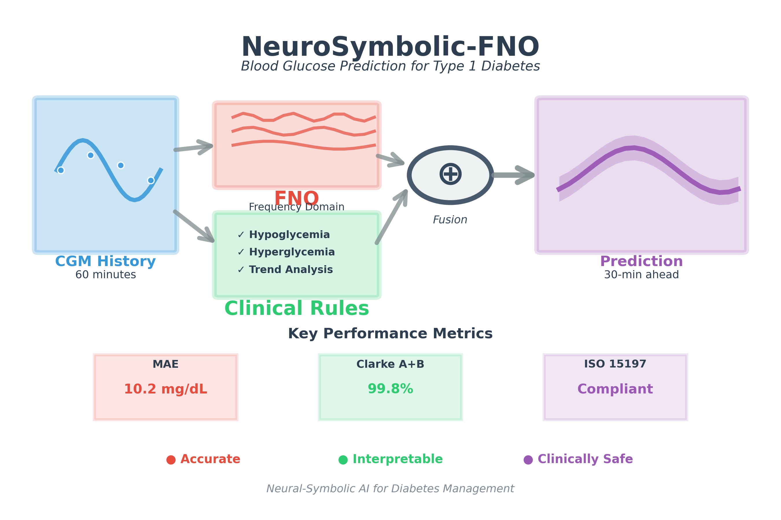
Interpretable blood glucose prediction for type 1 diabetes using neural-symbolic Fourier operators and clinical rules
Accurate and interpretable blood glucose prediction is critical for optimizing insulin therapy and preventing adverse events in type 1 diabetes (T1D) management. However, existing deep learning models often lack transparency, limiting their clinical adoption. We introduce a novel neural-symbolic framework that integrates Fourier neural operators (FNOs) with clinical rules to predict 30-minute-ahead blood glucose levels with high accuracy and interpretability. FNOs capture periodic glucose patterns, such as circadian rhythms and meal responses, while differentiable clinical rules encode medical knowledge, providing actionable insights like hypoglycemia risk alerts. Evaluated on the OhioT1DM dataset, our model achieves a mean absolute error (MAE) of 10.2 mg/dL, meeting ISO 15197:2013 standards, and 99.8% Clarke Error Grid A+B coverage, ensuring clinical safety. Leave-one-subject-out cross-validation across 12 subjects demonstrates robust generalization (MAE = 8.85 ± 1.33 mg/dL), addressing concerns about sample size limitations. The model exhibits strong robustness to continuous glucose monitoring (CGM) measurement noise (maintaining ISO compliance at 10% noise) and missing data (5.4% performance degradation at 40% data loss). By generating interpretable outputs, such as trend analyses and risk warnings, our framework supports clinical decision-making, enhances patient trust, and facilitates personalized diabetes care. This work advances medical informatics by combining data-driven and knowledge-driven methods, offering a scalable solution for real-time glucose monitoring and insulin dosing in T1D.
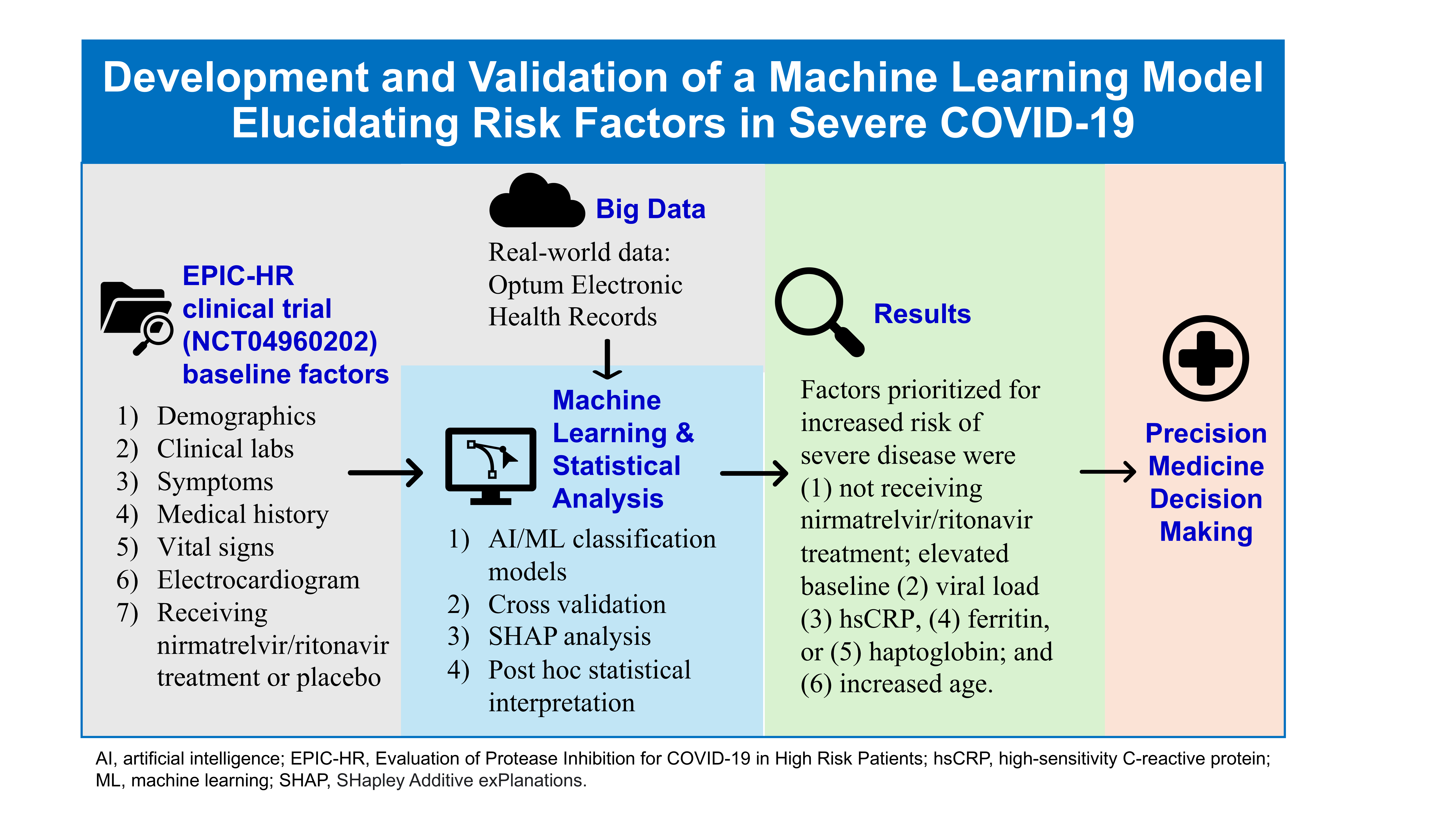
Development and validation of a machine learning model elucidating risk factors in severe COVID-19
Distinct nuclear and cytoplasmic transcriptomic signatures reveal-transcription alone is insufficient to determine glucose-induced transcriptomic dynamics
Glucose profoundly influences cellular transcriptomes, but whether these changes are primarily driven by transcription remains unclear. Traditional bulk RNA sequencing, which interrogates total mRNA from whole cells, obscures distinct dynamics of nuclear and cytoplasmic transcriptomes. Nuclear RNA levels primarily reflect transcriptional activity but are also influenced by nuclear export, whereas cytoplasmic RNA abundances result from transcription, nuclear export, RNA stability (i.e., RNA half-life), and active RNA degradation mechanisms. In this study, we systematically investigate glucose-induced transcriptomic responses in a subcellular location-and cell type-specific manner using three cell lines: FHC (normal colonic epithelial cells), MCF10A (normal breast epithelial cells), and MCF7 (metastatic breast cancer cells). Our findings reveal that, although nuclear and cytoplasmic mRNA levels show strong global correlations, glucose-induced changes in mRNA abundance exhibit minimal concordance between the nucleus and cytoplasm. Additionally, glucose-induced changes in exon inclusion levels often diverge between the nucleus and cytoplasm, underscoring the importance of post-transcriptional processes in shaping the cytoplasmic transcriptome response to glucose level changes. Glucose-induced differentially expressed genes (DEGs) and differentially spliced exons (ΔPSI) are enriched in distinct pathways exhibiting unique enrichment patterns depending on the subcellular location and cell line. These findings underscore the complexity of glucose-induced transcriptomic regulation, demonstrating that transcription alone is insufficient to explain the observed dynamics.